When performing a mixed dentition examination, the main goal is to determine whether there is need for interceptive orthodontic measures tha...
When performing a mixed dentition examination, the main goal is to determine whether there is need for interceptive orthodontic measures that will allow for the eruption of all the permanent teeth.
The earlier in the mixed dentition stage a problem is diagnosed and corrected, the better off patients will be as they continue to grow.
When performing an interceptive orthodontic examination, the following records are needed.
Records
1. Study Models
Study models are necessary because they allow you to evaluate the occlusion outside of the patient’s mouth. For example, abnormal wear patterns and crossbites can easily be seen.
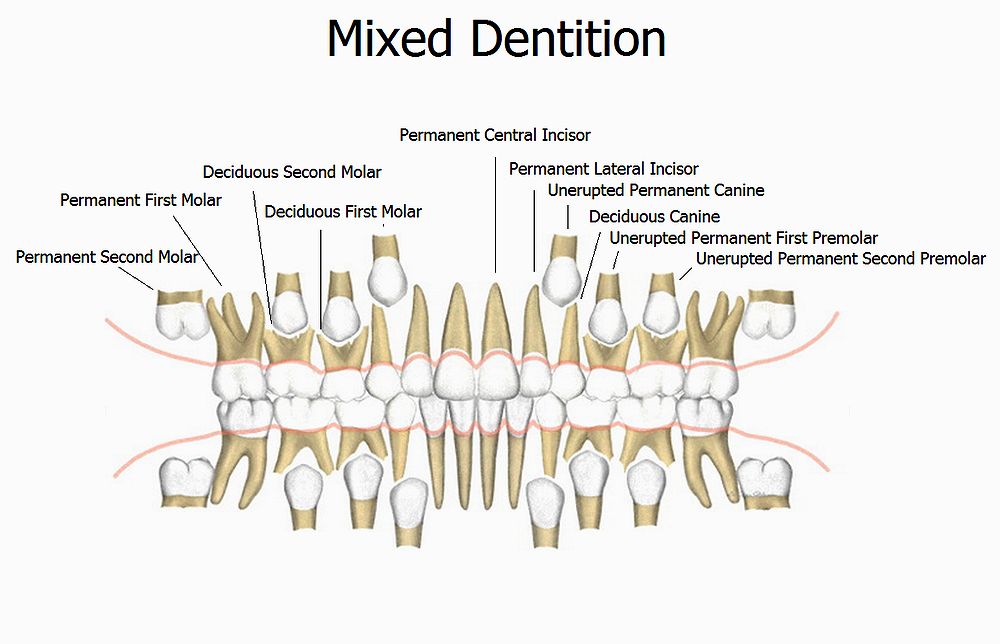
Study models also allow the practitioner to perform a mixed dentition analysis. Many mixed dentition analyses exist, such as the Tanaka and Johnston and Moyer’s prediction values. An accurate bite registration must also be taken as part of this record.
► Read Also : ORTHODONTIC : Guiding Unerupted Teeth into Occlusion: Case Report
2. Radiographs
2.1 Panoramic Radiograph
In the mixed dentition phase, the panoramic radiograph is useful for seeing permanent erupting teeth, crowding of teeth, space or lack of space between teeth, eruption paths, third molars, supernumerary teeth, and root apex formation (which is used to determine the patient’s dental age).
Using a panoramic radiograph is like seeing the world through a wide-angle lens, as compared to looking through a small looking glass, which could be considered analogous to full-mouth series of radiographs.
2.2 Lateral Head Film
(Cephalometric Radiograph)
Lateral head films are necessary when evaluating growing children to evaluate dentofacial proportions. As teeth erupt and growth occurs, the teeth relationships (within the jaws and skull) are part of a much bigger picture only visible with a cephalometric film and the appropriate cephalometric tracing.
In the mixed dentition, the following guidelines are designed to help in the decision process on when a cephalometric film is indicated.
Class II Patients:
Patients presenting with Class II dental relationships such as a distal step in primary second molars.
Patients with Class II relationships of permanent molars.
Patients who have a signi cant positive overjet and/or patients with mandibular retrusive profiles.
Class III Patients:
Patients with Class III relationships of permanent molars.
Patients who have a mesial step of primary second molars.
Patients who have a signi cant negative overjet (underbite).
Patients who have a protrusive profile of the mandible or retrusive profile of the maxilla.
Airway problems:
Airway problems diagnosed in children with open mouth breathing tendencies, such as turned up noses, allergic salute (wiping the nose with the hand in an upward swipe), or other medical history findings.
Vertical relationship problems:
Vertical relationship problems such as open bites associated with habits, airway problems, vertical skeletal growth problems, or patients with lip incompetency(lips do not touch or seal at mandibular rest).
Serial Lateral Head Films
Serial lateral head film radiographs are useful when monitoring growth in children with Class II or Class III tendencies, beginning at the first visit you diagnose them. They are also useful in comparing what orthodontically has really occurred after patients have been treated, by comparing pre- and posttreatment films.
3. Photographs
It is recommended that a full series of orthodontic photographs is taken for all patients. There is a proper way to take photographs, along with a way to retract soft tissues to capture vital anatomy, such as molar relationships.
The standard orthodontic photographs consist of eight pictures. Extraoral Photos: profile, frontal facial smiling, frontal facial at rest.
Intraoral Photos (teeth in occlusion): maxillary occlusal, mandibular occlusal, right and left buccal dental, and frontal dental.
There are other useful photos one can take when documenting an examination. For example, a patient with a tooth interference that causes a shift when intercuspation occurs can be documented by photographing the midlines at rest and with the teeth apart. When the patient occludes, the midlines will change, demonstrating the shift.
Close-up shots of individual teeth are also useful when documenting chips or decalcifications that you may be blamed for in the future after orthodontic treatment has been completed.
4. Other Records
Other records may also be needed, depending on the oral examination, such as anterior-posterior films (AP films) (for transverse analysis), conebeam 3-D imaging films (the new frontier in radiology), and/or occlusal films.
°Diagnosis Early Interceptive Orthodontic Problems - Part 1
°Michael Florman

No hay comentarios.